
We spoke with Dr. Francesco Neri, Director of Research and Operations at NeuroAge Therapeutics, San Francisco, a leading organization in the field of Alzheimer's disease research.
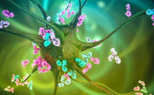
High-content imaging (HCI) has emerged as a powerful and versatile tool in Alzheimer's disease research, enabling scientists to investigate complex cellular behaviors and molecular alterations with exceptional detail and quantitative precision. HCI plays a critical role across various areas of research, including the development and analysis of cellular models, the study of amyloid-beta and Tau pathologies, high-throughput drug screening, and mechanistic investigations.
Additionally, it facilitates the exploration of advanced 3D culture systems and brain organoids, providing a more physiologically relevant context. By integrating high-resolution microscopy, automation, and sophisticated computational analysis, HCI generates comprehensive datasets that illuminate the intricate biological processes underlying Alzheimer’s disease.
Q: Tell us a little bit about yourself.
Dr. Neri: I gained my B.S. in biotechnology and my M.S. in pharmaceutical biotechnology at the University of Bologna in Italy. After that, I completed my PhD in the Campisi laboratory at the Buck Institute for Research on Aging, working on cellular senescence, a major driver of aging, and interventions for age-related diseases.
I’ve worked at NeuroAge Therapeutics since August 2024, developing our pre-clinical drug development strategy and leading clinical operations for our NeuroAge Test.
Q: Can you tell us about your company and your focus areas?
Dr. Neri: NeuroAge Therapeutics is a pioneering longevity biotech startup dedicated to rejuvenating minds and revolutionizing brain health. The company has an innovative approach to combat neurodegenerative disorders like Alzheimer's by reversing brain aging. We’re achieving this by leveraging human omics and AI to identify targets causally implicated in brain aging.
At the same time, we’re providing the NeuroAge Test, the most comprehensive brain health assessment available to date. By tracking people’s brain health and biomarkers earlier in life, we are making preventative interventions a reality.
Q: What are you using high-content imaging technology for?
Dr. Neri: We culture neuronal cells to study the processes of brain aging and to test for potential therapeutics against aging. Cell imaging is a crucial technology for us, we stain for certain proteins or markers and then using the Operetta CLS™ high-content analysis system we do high-throughput imaging to gather large amounts of image data to answer the questions that we’re asking.
For example, we assess the neuroprotective capabilities of our compounds by exposing cultures to aging/disease-associated stressors and the compounds. We then use the Operetta CLS live-cell imaging capabilities to measure cell death over time, followed by staining for neuronal health markers at the end of treatment as end-point measurements. The Operetta CLS makes it easy and convenient to generate large amounts of rich data from individual experiments that are easily reproducible.
Q: How has high-content imaging technology helped to answer your research questions?
Dr. Neri: High-content imaging allows us to gather a lot of data really quickly – it would be much more time-consuming to do this by other methods. With the Operetta CLS system, we can speed things up considerably, which also makes the overall process less expensive.
Q: What aspects of the Operetta CLS have been particularly helpful in your work?
Dr. Neri: The Operetta CLS system is fast and the software is very intuitive - both for setting up the imaging acquisition as well as creating image analysis pipelines. And having the Revvity technical support team on hand to help when we have any questions or issues with assay development is very helpful too.
Q: What are your plans for this research in the future, and what are you most excited about?
Dr. Neri: We’re excited to explore the brightfield segmentation feature of the Operetta CLS to expand our multiplexing capabilities. By using brightfield for cell segmentation, we can free up a fluorescent channel and allocate it to additional markers, enhancing the depth and scope of our analyses.
The Operetta CLS enables efficient, high-quality imaging for advanced research applications. Researchers interested in exploring how high-content imaging could provide deeper insights into the biology being studied are encouraged to contact Revvity to learn more.