


Cytopathic effects of PI3 virus on Vero cells measured by confluence percentages
In this experiment, the goal is to measure the time-course CPE of PI3 virus on the Vero host cells in order to determine the earliest time point that CPE can be automatically detected.
The Vero host cells are seeded in 6-well microplates, incubated and allowed to adhere overnight. The host cells are inoculated with PI3 virus as the positive sample and media for the negative control. The 6-well microplates are scanned at 10% of the well that required less than 1 min/plate and analyzed using Celigo™ image cyotmeter
 Celigo Image Cytometer
from day 0 to 12. The Vero cell confluence percentages are measured directly in brightfield imaging to determine the CPE.
Celigo Image Cytometer
from day 0 to 12. The Vero cell confluence percentages are measured directly in brightfield imaging to determine the CPE.
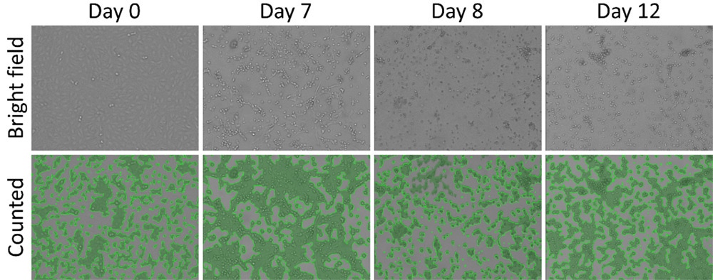
Time-course brightfield and analyzed images (Confluence) for the positive PI3 virus. The reduction in confluence % of Vero cells indicates cytopathic effects.
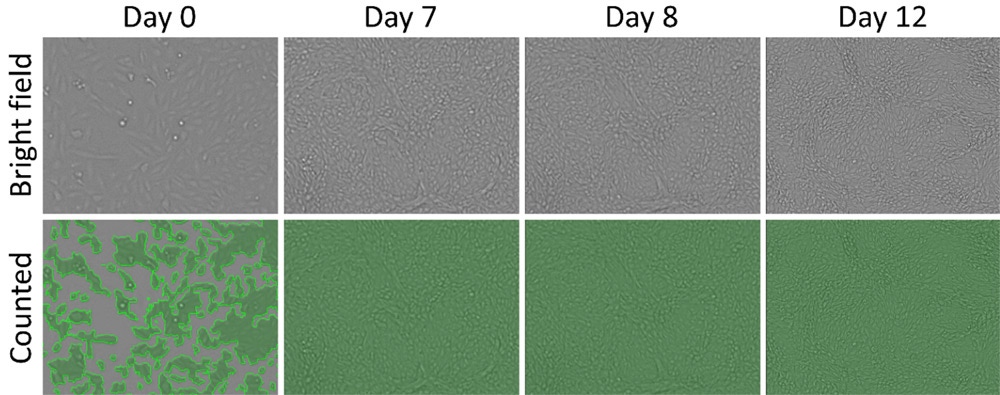
Time-course brightfield and analyzed images (Confluence) for the negative media control.
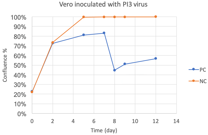
Time-course confluence percentage results, showing the CPE occurring on day 5.
Example CPE measurement performed using the Celigo image cytometer:
You may also be interested in these products


Part number:
200-BFFL-5C

Part number:
CS2-0106-5ML,
CS2-0106-25ML
List price:
USD 133.00 - USD 594.00
Log in to view
your online price
")
List price:
USD 286.00 - USD 5,260.00
Log in to view
your online price
")
List price:
USD 286.00 - USD 5,260.00
Log in to view
your online price
of 5
For research use only. Not for use in diagnostic procedures.