


Cytopathic effects of measles virus on MRC-5 cells measured by confluence percentages
In this experiment, the goal is to measure the time-course CPE of the measles virus on the MRC-5 host cells in order to determine the earliest time point that CPE can be automatically detected.
The MRC-5 host cells are seeded in 6-well microplates, incubated and allowed to adhere overnight. The host cells are inoculated with the measles virus as the positive sample and media for the negative control. The 6-well microplates are scanned at 10% of the well that required less than 1 min/plate and analyzed using Celigo™ image cyotmeter
 Celigo Image Cytometer
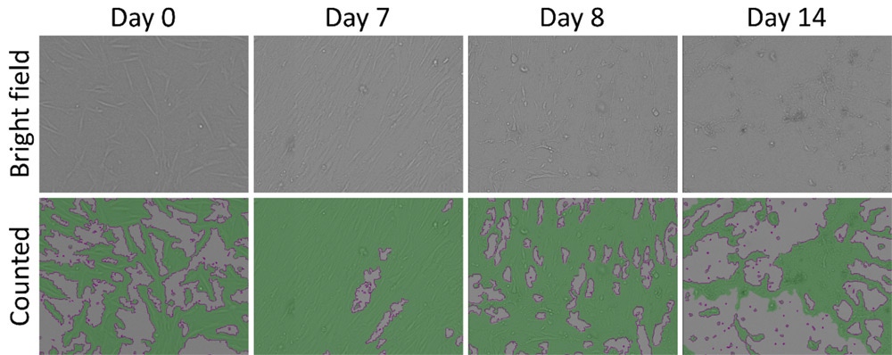
from day 0 to 14. The MRC-5 cell confluence percentages are measured directly in bright field imaging to determine the CPE.
Celigo Image Cytometer
from day 0 to 14. The MRC-5 cell confluence percentages are measured directly in bright field imaging to determine the CPE.
Time-course brightfield and analyzed images (Confluence) for the positive measles virus. The reduction in confluence % of MRC-5 cells indicates cytopathic effects.
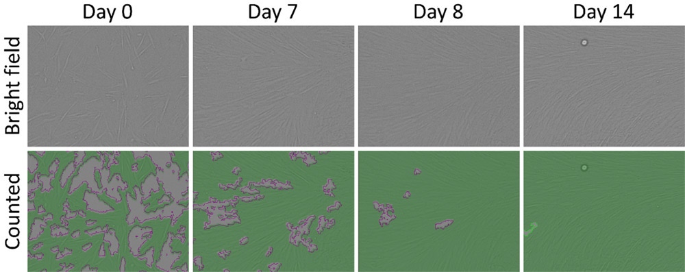
Time-course brightfield and analyzed images (Confluence) for the negative media control.
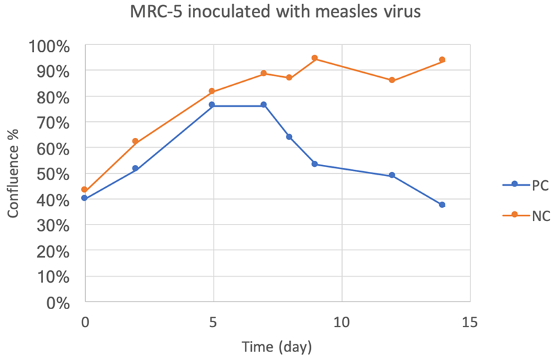
Time-course confluence percentage results, showing the CPE occurring on day 8.
You may also be interested in these products


Part number:
200-BFFL-5C

Part number:
CS2-0106-5ML,
CS2-0106-25ML
List price:
USD 133.00 - USD 594.00
Log in to view
your online price
")
List price:
USD 286.00 - USD 5,260.00
Log in to view
your online price
")
List price:
USD 286.00 - USD 5,260.00
Log in to view
your online price
of 5
For research use only. Not for use in diagnostic procedures.