
Therapeutic development has relied on cell culture and animal models to evaluate the potential safety and efficacy of treatments. Studying disease models or treatments in two-dimensional (2D) cell culture is limited because these models do not fully capture the complexities of three-dimensional (3D) biological systems. In addition, animal-based drug discovery has faced challenges in translating preclinical findings into clinical studies, leading to high failure rates.
Developing methods to bridge the gaps between conventional 2D cell culture and living organisms has led to increasing use of 3D cell culture models. 3D modeling has been useful for studying disease biology and evaluating cellular responses to experimental treatments.1 Many have used the terms organoids, spheroids, and 3D culture interchangeably; however, these are distinct terms that differ in how they are cultured, maintained, and studied.
Key takeaways:
- The key differences between spheroids and organoids, and how their characteristics influence their use in 3D culture research.
- The advantages and limitations of commonly used techniques for analyzing 3D cultures, including plate readers, flow cytometry, sequencing, and imaging.
- How imaging-based approaches can be used to monitor 3D cultures over time and support analysis of growth, morphology, and cellular responses.
What are the differences between spheroids and organoids?
Spheroids are spherical cellular units that are cultured as aggregates. They are derived from cell lines, primary cells, and tumor cells. In the appropriate conditions, they self-assemble into heterogeneous layers that include proliferating quiescent and necrotic cells. Spheroids are relatively easy to culture and can be used for large drug screenings, as well as for cell migration and invasion studies. However, spheroids do not have a complex structure, don’t always resemble original tissue, and have limitations for long term studies.1
Organoids are spherical cellular units derived from embryonic stem cells, adult stem cells, or iPSCs, and can also be generated from tumor cells and tissues. Organoids self-organize and assemble via differentiation of cells, and they require physical and chemical cues. Organoids develop into more complex structures with different lineages that are more like organs. Organoids are more difficult to grow and maintain since they require extracellular matrix (EM) and growth factors. Their formation can take months, and they can be preserved for long-term studies. Organoids are useful for studying genetic pathways, disease models, cancer, and development. Patient derived organoids (PDOs) are formed from patient cells and are used in research to investigate disease biology and responses to experimental treatments.1
Analysis of 3D cultures
Once researchers can successfully grow and maintain 3D cultures, analysis tools are required to determine treatment responses in disease or cancer models. Table 1 summarizes the current techniques with the pros and challenges for each technique. Typically, after treatment with multiple doses and conditions, researchers compare the endpoint of the 3D culture with the initial timepoint. A plate reader can track changes in fluorescent proteins and is easy to scale up in 96- or 384-well plates to test multiple conditions. This is helpful for screening drug responses; however, normalization based on cell number is required, and this can only be achieved as an endpoint assay.
Flow cytometry is another tool that provides detailed information about the cell composition of 3D cultures through multiplexing and analysis. Flow cytometry requires expert-level knowledge, and dissociation of the 3D culture is required, making it an endpoint assay. Single-cell sequencing is another technique that provides insight and is common for PDOs. Genetic pathways and patient-specific information can be obtained through sequencing, which can help uncover the molecular mechanisms associated with treatment. These are all tools to monitor 3D culture at the endpoint, meaning that kinetic analysis is not readily possible to track growth and response to drugs in real time.
| Technique | Pros | Challenges |
|---|---|---|
| Plate reader | Fast, easy, large scale capable, whole well read out | Normalization required, limited insight, endpoint |
| Flow cytometry | Detailed data, multiplexing and flexible, powerful analysis | Complex, dissociate cells, expert user |
| Single cell sequencing | Powerful analysis, genetic pathways discovered, personalized medicine, molecular mechanism uncovered | Large datasets can be cumbersome to interpret, endpoint assay |
| Imaging-confocal microscopy | Single-cell data insight without disturbing cells, kinetic assay, straightforward data | Analysis can be challenging, time consuming, expert user |
Imaging 3D cell cultures
To monitor 3D culture over time, imaging-based approaches are commonly used. This is a challenge since spheroids and organoids can be large, involve multiple focal planes, and imaging requires time for image acquisition and analysis. To obtain high-quality images at several Z positions, confocal microscopy can be used to detect size changes, numbers, and fluorescent proteins or cells over multiple time points. This is because imaging can be conducted multiple times throughout the experiment, since disrupting the structure is not required. However, this is time consuming, and analysis can be challenging without the right tools.
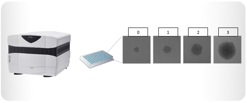
To improve imaging methods, Revvity offers several solutions to automate imaging and analysis steps. The CeligoTM image cytometer is a plate-based, multichannel brightfield and fluorescent system that automatically images and analyzes 3D culture. It can acquire whole-well images of a 96-well plate and analyze a plate in approximately five minutes. It can provide the number of spheroids or organoids, their sizes, and fluorescent signals in a single assay. Due to its speed, changes in growth can be detected over time (Figure 1).
Figure 1: The Celigo image cytometer can scan and analyze plates to detect 3D culture over the course of several days and track morphological changes.
Since the Celigo image cytometer acquires whole-well imaging, it can also detect multiple organoids for screening purposes (Figure 2). Revvity’s Image Artist™ is another tool to analyze organoids and can be combined with images acquired from the Celigo image cytometer, supporting Z-stack image analysis. Image Artist can convert images acquired on the Celigo image cytometer into maximum intensity projections (MIP).
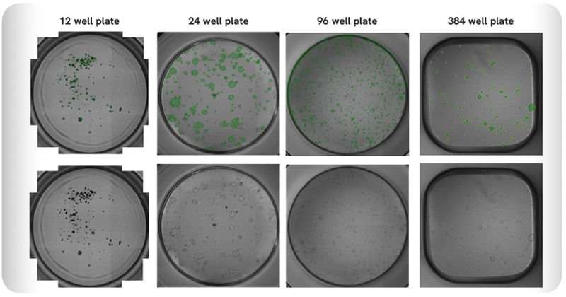
Figure 2: The Celigo image cytometer can rapidly scan and analyze organoids on multiple plate formats. Counted PDOs are shown in green pseudo-color.
The Celigo image cytometer is a useful tool for rapid imaging and analysis and for detecting changes over time in response to treatment. For detailed imaging at single-cell resolution in 3D, other options are to include the Opera PhenixTM or OperettaTM CLS high-content screening systems. These instruments provide confocal image quality and enhanced image analysis tools once a drug treatment or dosage is chosen with the Celigo image cytometer.
To learn more about the Celigo image cytometer and 3D culture, visit our dedicated webpages.
Reference:
- Gunti S, Hoke ATK, Vu KP, London NR Jr. Organoid and Spheroid Tumor Models: Techniques and Applications. Cancers (Basel). 2021 Feb 19;13(4):874. doi: 10.3390/cancers13040874. PMID: 33669619; PMCID: PMC7922036.
For research use only. Not for use in diagnostic procedures.