


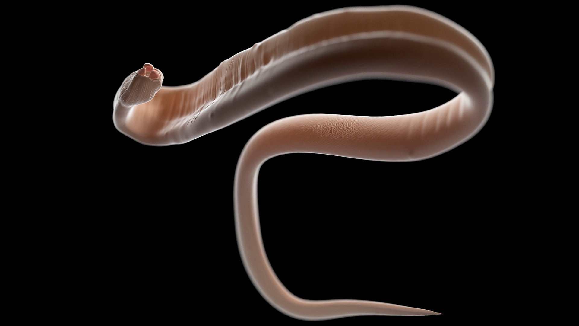

MicroRNAs (miRNAs) are central regulators of gene expression in animals, with well-characterized roles in development, immunity, and disease in humans. In parasitic helminths (nematodes and platyhelminths), miRNAs are similarly abundant and developmentally regulated, controlling processes such as stage transitions, reproduction, and host adaptation1.
Over the last decade, it has become clear that many helminths secrete miRNAs into the host environment, frequently packaged within extracellular vesicles (EVs). A landmark study demonstrated that a nematode parasite secretes exosome-like vesicles carrying parasite small RNAs (including miRNAs) that can enter mammalian cells and modulate host responses, providing a mechanistic foothold for cross-species RNA transfer2.
Extracellular vesicles are lipid-bounded particles released by cells that transport proteins, lipids, and nucleic acids. Helminth-derived EVs have now been isolated from multiple species and life stages, and their RNA cargo has been profiled. Comparative synthesis of the field highlights both shared and clade-specific EV miRNA features across nematodes, trematodes, and cestodes, while also emphasizing methodological heterogeneity across studies3.
These findings support a paradigm in which helminth miRNAs participate in molecular exchange between parasite and host, though the strength of evidence differs depending on whether the claim concerns secretion and detectability (strong) versus in vivo gene-regulatory function (still emerging)1.
Progress in this field has been driven primarily by improvements in small RNA sequencing, EV purification protocols, and bioinformatic pipelines that distinguish parasite reads from host background and apply conservative criteria for miRNA annotation in non-model organisms. Reviews of helminth EV cargo repeatedly stress that differences in EV purification and RNA profiling workflows can shape reported miRNA content and cross-study comparability3.
Functional evidence and diagnostic implications
Small-RNA sequencing studies show a non-random miRNA composition within helminth-derived EVs, frequently featuring conserved families (e.g. let-7 and others), alongside lineage- and stage-specific miRNAs. In liver fluke biology, for example, specific miRNAs have been reported to be enriched in EV relative to whole-parasite RNA, suggesting active sorting rather than passive release4. Importantly, EV encapsulation is widely discussed as a mechanism that protects extracellular RNAs from degradation, contributing to their stability in host-facing environments and increasing their likelihood of biological activity1,3.
Helminth-derived EVs have shown to be internalized by mammalian immune cells in vitro. In a well-cited study, EVs from the gastrointestinal nematode Heligmosomoides polygyrus were taken up by macrophages and supressed classical activation pathways, an effect attributed to the combined action of EV-associated proteins and small RNAs5. Subsequent work in Trichinella spiralis, demonstrated that specific EV-associated miRNAs (including let-7-5p) can modulatemacrophage polarization in cultured cells through target-linked assays, supporting a direct, sequence-dependent regulatory effect.6 Complementary studies in epithelial models indicate that helminth EV miRNAs (such as miR-153) can influence host cell survival pathways, reinforcing the concept that miRNAs constitute functional cargo under defined experimental conditions7.
Beyond potential functional roles, helminth-derived miRNAs have attracted substantial interest as diagnostic biomarkers. In schistosomiasis, multiple studies have detected parasite-derived miRNAs in host circulation and evaluated their association with infection status, parasite burden, and disease progression8,9. These findings suggest that parasite miRNAs (freely circulating or EV-associated) complement existing diagnostic approaches. However, reviews of the field constantly emphasize current limitations related to sensitivity, early-stage detection, and cross-study variability, underscoring the need for standardized sampling, RNA profiling, and analytical frameworks1,3.
Conclusions
Definitive demonstrations of miRNA-specific effects in vivo are still scarce. As a result, the magnitude, physiological relevance, and precise mechanisms of parasite miRNA activity during natural infections remain uncertain. Interpretation is further complicated by the complex composition of helminth EVs and by variability in EV isolation and RNA profiling methodologies across studies.
From a translational perspective, the detectability of parasite-derived miRNAs in host biofluids supports their potential as non-invasive biomarkers of infection and treatment response. However, therapeutic applications remain speculative and will require stronger mechanistic and in vivo validation. Overall, helminth-derived miRNAs highlight small RNAs as components of host–parasite communication.
References:
- Mu, Y., et al. (2021). Parasitic Helminth-Derived microRNAs and Extracellular Vesicle Cargos as Biomarkers for Helminthic Infections. Front Cell Infect Microbiol. 11:708952. doi: 10.3389/fcimb.2021.708952.
- Buck, A.H., et al. (2014). Exosomes secreted by nematode parasites transfer small RNAs to mammalian cells and modulate innate immunity. Nat Commun. 5:5488. doi: 10.1038/ncomms6488.
- Sotillo, J., et al. (2020). The protein and microRNA cargo of extracellular vesicles from parasitic helminths - current status and research priorities. Int J Parasitol. 50(9):635-645. doi: 10.1016/j.ijpara.2020.04.010.
- Fromm, B., et al. (2015). The revised microRNA complement of Fasciola hepatica reveals a plethora of overlooked microRNAs and evidence for enrichment of immuno-regulatory microRNAs in extracellular vesicles. Int J Parasitol. 45(11):697-702. doi: 10.1016/j.ijpara.2015.06.002.
- Coakley, G., et al. (2017). Extracellular Vesicles from a Helminth Parasite Suppress Macrophage Activation and Constitute an Effective Vaccine for Protective Immunity. Cell Rep. 19(8):1545-1557. doi: 10.1016/j.celrep.2017.05.001.
- Liu, Y., et al. (2025) miRNA let-7-5p present in the extracellular vesicles of Trichinella spiralis newborn larvae inhibits the function of M1-type RAW264.7 macrophages by targeting C/EBPδ. Parasites Vectors 18, 199 (2025). doi:10.1186/s13071-025-06802-2.
- Wang, R., et al. (2023). Exosome-delivered miR-153 from Trichinella spiralis promotes apoptosis of intestinal epithelial cells by downregulating Bcl2. Vet Res. 54(1):52. doi: 10.1186/s13567-023-01186-6.
- Cai, P., et al. (2015). Circulating miRNAs: Potential Novel Biomarkers for Hepatopathology Progression and Diagnosis of Schistosomiasis Japonica in Two Murine Models. PLoS Negl Trop Dis. 9(7):e0003965. doi: 10.1371/journal.pntd.0003965.
- Mu, Y., et al. (2020). Parasite-derived circulating microRNAs as biomarkers for the detection of human Schistosoma japonicum infection. Parasitology. 147(8):889-896. doi: 10.1017/S0031182019001690.
For research use only. Not for use in diagnostic procedures.


